Authors: Jade Harkin, Cátia Gomes, Reham Afify, and Jason S. Meyer
PNAS, 19 August 2026
Lux imaging captures dynamic microglial phagocytosis alongside retinal ganglion cell degeneration.
Optic neuropathies such as glaucoma involve progressive degeneration of retinal ganglion cells, but the contribution of human microglia and astrocytes to retinal ganglion cell health and disease remains difficult to study. In this work, researchers developed human pluripotent stem cell-derived (hiPSC) co-culture and tri-culture systems combining retinal ganglion cells, microglia, and astrocytes to examine how these cell types interact under homeostatic and inflammatory conditions.
Under homeostatic conditions, microglia enhanced retinal ganglion cell growth and maturation, increasing neurite length and complexity as well as neuronal firing and bursting activity. When microglia were driven into a pro-inflammatory state, retinal ganglion cells showed reduced soma size, neurite growth, firing rate, and bursting activity, while astrocytes adopted a more reactive phenotype.
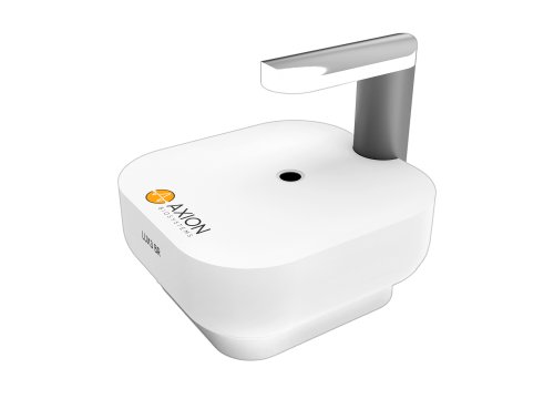
To characterize microglial function directly, the researchers used Axion BioSystems’ Lux live-cell imaging system to track phagocytosis in control and LPS-activated microglia over 12 hours. Lux imaging revealed significantly greater phagocytic activity in activated microglia, adding a kinetic functional readout to the model. Together with electrophysiology, morphology, and single-cell RNA sequencing, these data provide a more complete view of how neuroinflammatory cell interactions contribute to retinal ganglion cell degeneration in glaucoma and other optic neuropathies.