
Organoid Analysis Module
Automate organoid counting, monitoring, and morphological analysis
Track organoid formation, growth, and morphology over time with automated whole-vessel imaging and AI-driven analysis. The Organoid Analysis Module for Omni identifies, counts, and measures organoids across supported culture vessels, helping you replae manual counting with a more standardized, reproducible workflow.
Key Features
Key Features

Count every organoid across the vessel
Whole-vessel imaging captures all organoids to reduce sampling bias.

Monitor development over time
Automatically image cultures at defined intervals to track formation, growth, and morphology longitudinally.

Reduce manual variability
Replace time-consuming, user-dependent counting with automated analysis.

Quantify morphology
Measure organoid count, diameter, area, roundness, and darkness to compare populations and conditions.
Stop counting organoids by hand
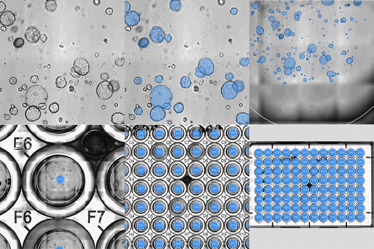
Manual orgnaoid counting can be time-consumig and inconsistent, especially as experiments scale. The Organoid Analysis Module automatically detects and measures organoids across the entire culture vessel, so you can compare conditions, generate population distributions, and follow development over time without manually reviewing each well.
Overview
What can the Organoid Analysis Module measure?

Organoid count
Track the number of detected organoids over time.

Diameter
Measure changes in organoid size during formation and growth.

Area
Quantify two-dimensional projected organoid area.

Roundness
Compare changes in organoid morphology and shape.

Darkness
Track image-intensity characteristics as organoids develop.
Whole-vessel imaging for more representative data
Omni can image the entire supported culture vessel at each timepoint, helping you capture more of the organoid population and reduce field-of-view bias.
- ✓ Image every organoid in the vessel
- ✓ Follow the same culture repeatedly
- ✓ Identify changes between manual observations
Standardize organoid monitoring from formation through maturation
>> Early formation
Track embryoid body and organoid number, size, and shape as cultures establish.
>> Growth and morphology
Monitor individual organoids and population changes over time.
>> Compare conditions
Evaluate media, matrices, treatments, or differentiation protocols.
>> Build longitudinal datasets
Generate sequential visual and quantitative records without removing cultures from the incubator.
From images to quantitative results
Omni combines automated image acquisition with cloud-based analysis to turn large organoid imaging datasets into quantitative results.
Automatic detection
Population analysis
Growth trajectories
Review images and data
Frequently asked questions about the Organoid Analysis Module
The Organoid Analysis Module automatically detects, counts, and measures organoids in supported culture vessels. It helps researchers monitor organoid formation, growth, and morphology over time without relying on manual counting.
The module can quantify organoid count, diameter, area, roundness, and darkness. These measurements can be used to compare populations, culture conditions, and changes over time.
Yes. Omni can repeatedly image the same culture at defined intervals, allowing researchers to follow organoid formation, growth, and morphological changes longitudinally.
When properly configured, the module largely eliminates manual counting by automatically detecting and measuring organoids across the imaged culture area. Whole-well images are saved to the portal, making it easy to manually verify the automated counts if needed.
Yes. Whole-vessel imaging captures the full organoid population rather than selected fields of view, which can reduce sampling bias and provide a more representative dataset.
Yes. Researchers can compare organoid number, size, morphology, and growth across different media, matrices, differentiation protocols, treatments, or other experimental conditions.
Yes. The Organoid Analysis Module can track early organoid or embryoid body formation and monitor changes in population and morphology as cultures mature. For iPSC-derived organoids, the iPSC Analysis Module can also support earlier stages of the workflow, enabling continuous monitoring from 2D iPSC culture through 3D organoid development.
The module can analyze brightfield images, allowing organoid growth and morphology to be monitored without fluorescent labeling for routine counting and morphology measurements.
Automated detection applies the same analysis approach across images and timepoints, helping reduce variability introduced by manual counting, subjective field selection, and differences between users.
For Research Use Only. Not for use in diagnostic procedures.


