AUTHORS: Rui Zhang, Xinbo Ji, Han Yu, Jingwen Xu, Yu Wang, Ping Sun, Yingxin Wang, Yao Tang, Zexin Zhan, Yichang Jiao, Didi Shan, Pengfei Lin, Dong-dong Wang, Yuying Zhao, Xianyang Liu, Chuanzhu Yan, Jianqiao Li, Mingfeng Li, Fuchen Liu, and Shengping Hou
CNS Neuroscience & Therapeutics, 03 August 2026
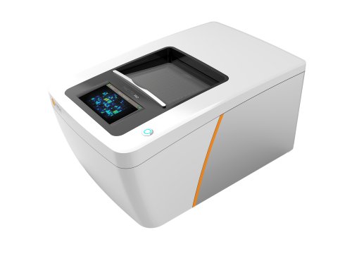
Axion MEA recordings reveal reduced electrophysiological activity in patient-derived retinal organoids with Usher syndrome 3A.
Usher syndrome type 3A (USH3A) causes progressive hearing loss and retinitis pigmentosa, but the mechanisms driving retinal degeneration remain incompletely understood. In this study, researchers generated patient-derived retinal organoids to investigate how CLRN1 variants affect retinal cell function and survival. Single-cell RNA sequencing showed that CLRN1 expression was concentrated in Müller cells, pointing to an important role for these retinal glia in disease progression.
The team found that CLRN1 variants disrupted mitochondrial function in Müller cells, leading to secondary mitochondrial impairment, oxidative stress, and apoptosis in photoreceptors. The retinal organoids also showed reduced photoreceptor marker expression and increased cell death. Using an Axion MEA system, researchers measured spontaneous electrical activity in Day 150 retinal organoids and found significantly lower weighted mean firing rates and spike counts in USH3A organoids compared with healthy controls.
The researchers also evaluated idebenone, a mitochondrial-targeted antioxidant, and found that treatment partially improved mitochondrial respiration and related functional deficits. Together, the findings identify Müller cell mitochondrial dysfunction as a potential driver of retinal degeneration in USH3A and demonstrate how patient-derived retinal organoids can connect molecular disease mechanisms with functional electrophysiological changes.